Figure 1.
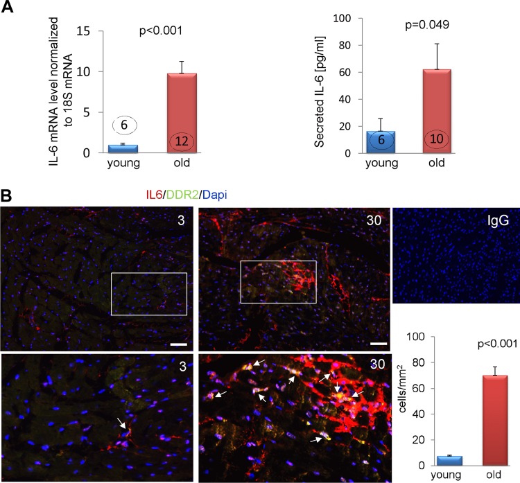
The expression of IL-6 in cardiac mesenchymal fibroblasts is affected by aging. A) Expression of IL-6 in quiescent fibroblasts derived from young and old MSCs were analyzed by quantitative PCR and by ELISA, 24 h after the cell cycle was synchronized. B) Double immunofluorescence labeling of IL-6 (red) and DDR2 (green) in 3- and 30-mo-old mouse hearts. Double positive cells are visualized in yellow/orange color. Middle panel shows magnified image of the section marked by white squares. Bottom panel shows morphometric analysis of IL-6+DDR2+ cells in heart sections. Nuclei were stained with DAPI. Scale bar, 20 μm. P < 0.05 denotes statistical significance. The number in each column corresponds to the number of donor animals from which cells were derived for these experiments except for immunofluorescence staining where sections from 3 animals per age-group were analyzed. Young or 3 signifies cells or hearts derived from 3-mo-old mice and old or 30 denotes cells or hearts obtained from 30-month old mice. Arrows point to double positive IL6+DDR2+ fibroblasts.

