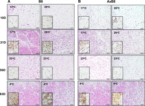
Figure 4.

Histologic examination of inguinal BAP. Sections of ING from 10, 21, 56 (moved to 23°C at weaning), and 63D (moved to 23°C at weaning and exposed to 4°C for 7 days) B6 (C57BL/6J) (A) and AxB8 (B) mice maintained at 17 or 29°C during the preweaning period. Large images show the complex patterns of reversible white and brown adipocyte morphology as a function of age and ambient temperature, stained with hematoxylin and eosin. Scale bar, 100 µm. Inset images show anti-UCP1 immunohistochemical staining, counterstained with hematoxylin. Scale bar, 50 µm.
