Figure 2.
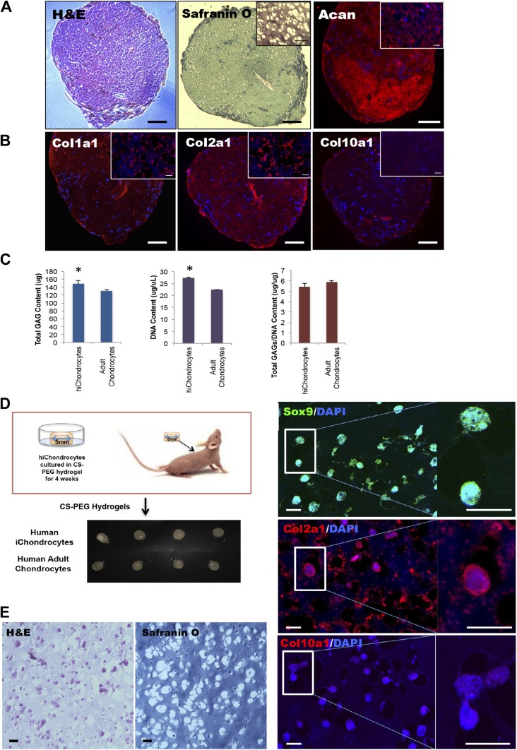
A–C) In vitro and (D–F) in vivo cartilage generation by hiChondrocytes. A) Representative hematoxylin and eosin (H&E), Safranin O, and (Acan) staining. B) Representative immunofluorescence staining for Col1a1, Col2a1, and Col10a1 in hiChondrocyte-derived cell pellets. C) Quantification of GAG production in hiChondrocyte-derived cell pellets in vitro. Data are presented as means ± sd of 3 independent experiments. *P < 0.05. D) hiChondrocytes encapsulated in CS-PEG hydrogels and engrafted subcutaneously in immunodeficient mice. E) Representative H&E and Safranin O staining of hydrogel-capsulated hiChondrocytes cultured for 4 wk in vivo. F) Confocal microscopy for representative immunofluorescence staining for chondrocyte markers, Sox9, Col2a1, and Col10a1 in hydrogel capsulated hiChondrocytes cultured for 4 wk in vivo. Scale bars, 100 μm; (B) inset 20 μm.

