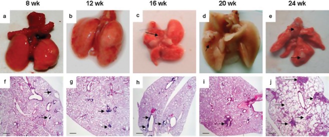
Figure 1.

Tumor pathology in mouse lungs with Cul4A overexpression. (a) Wild-type mouse lungs eight weeks post-infection with Ade-green fluorescent protein. The surface of the lungs is smooth and uniform. (b) Lox-Cul4A mouse lungs 12 weeks post-infection with adenovirus expressing Cre-recombinase (AdenoCre). The surface of the lungs is also smooth and uniform. (c) Lox-Cul4A mouse lungs 16 weeks post-infection with AdenoCre. The surface of the lungs have a bumpy appearance, arrows indicate the lesions. (d) Lox-Cul4A mouse lungs 20 weeks post-infection with AdenoCre. An isolated cobblestone-like nodule on the surface of the lungs is visible; the arrow indicates the lesion. (e) Lox-Cul4A mouse lungs 24 weeks post-infection with AdenoCre. Several nodules on the surface of the lungs have appeared; arrows indicate the lesions. (f) Histological sections of wild-type mouse lungs eight weeks post-infection with AdenoCre. (g) Histological sections of Lox-Cul4A mouse lungs 12 weeks post-infection with AdenoCre; the arrow shows a single lesion. (h) Histological sections of Lox-Cul4A mouse lungs 16 weeks post-infection with AdenoCre; arrows indicate isolated lesions. (i) Histological sections of Lox-Cul4A mouse lungs 20 weeks post-infection with AdenoCre; arrow shows a single adenocarcinoma-like lesion. (j) Histological sections of Lox-Cul4A mouse lungs 24 weeks post-infection with AdenoCre; arrows show diffuse severe adenocarcinoma-like lesions. Scale bar indicates 200 μm. Wk, weeks.
