Figure 2.

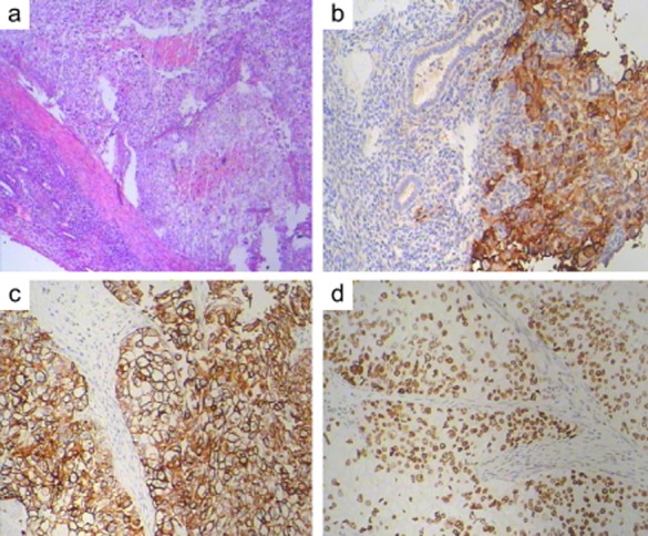
Pathological examination of the mass in the uterus, immunohistological staining for epidermal growth factor receptor, cytokeratin-7, and P53.

Pathological examination of the mass in the uterus, immunohistological staining for epidermal growth factor receptor, cytokeratin-7, and P53.