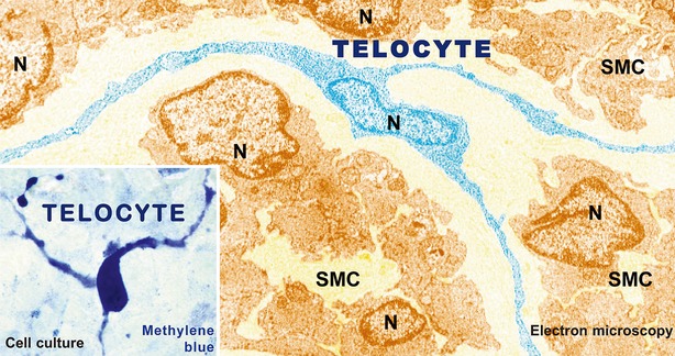
Figure 1.

Representative electron microscopy image of a telocyte. A telocyte (TC) with at least three prolongations with several ‘beads’ along telopodes (Tps) is digitally coloured in blue. SMC: smooth muscle cell; N: Nuclei. Original magnification ×6800. Reproduced with permission from 8.
