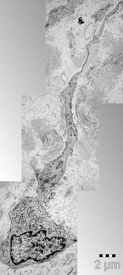
Figure 2.

Three sequenced electron microscopy images of a telocyte. A telocyte (TC) with typical long and thin telopode (Tp) extending from the cell body; scale bars: 2 μm. Courtesy of Dr. LM. Popescu, Department of Ultrastructural Pathology, Victor Babeş National Institute of Pathology, Bucharest, Romania.
