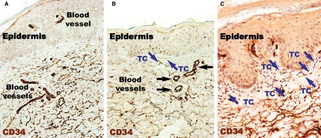
Figure 3.

CD34 immunohistochemistry of a psoriatic plaque (A) revealing a lower density of positive expression in the papillary dermis compared to distant uninvolved skin (B). However, the papillary dermis of treated skin has a density comparable to uninvolved skin (C). CD34 expression is higher within the reticular dermis of psoriatic skin (A) and treated skin (C) compared the reticular dermis of normal skin. Silhouettes of CD34-positive telocytes (TC; blue arrows) with long telopodes were observed in the papillary dermis of normal skin (A) and treated skin (C). Magnification 200×. Black arrows indicate blood vessels.
