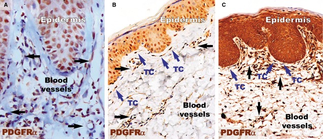
Figure 5.

PDGFRα immunohistochemistry revealed an increased density of PDGFRα-positive structures in the papillary dermis of a psoriatic plaque (A) and treated skin (C) compared to the papillary dermis of distant uninvolved skin (B). (A) The papillary dermis of the lesional skin has increased cellularity, but no PDGFRα-positive telocytes were observed. Blood vessels (black arrows) have dilated diameters within the dermis. (B) Telocytes (TC; blue arrows) positive for PDGFRα are seen in the papillary dermis of uninvolved skin. TCs have very long prolongations that run parallel to the basement membrane of the epidermis. (C) PDGFRα-positive telocytes (TC; blue arrows) are observed among the increased cellularity of the papillary dermis of treated skin and have the same orientation as in uninvolved skin. The blood vessels have comparable diameters to those in uninvolved skin. Black arrows indicate blood vessels. Magnification 200×.
