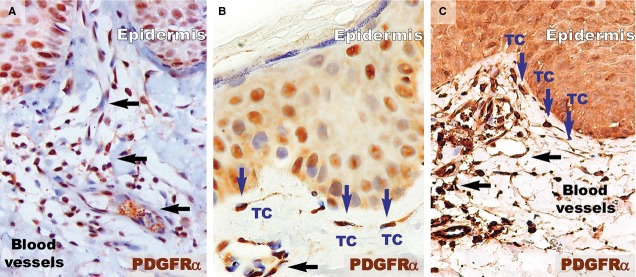
Figure 6.

Higher magnifications of immunohistochemistry for PDGFRα in the papillary dermis show the absence of telocytes but the presence of inflammatory cells (mainly lymphocytes) in a psoriatic plaque (A). However, in the distant uninvolved skin (B), telocytes (TC, blue arrows) with very long telopodes are situated in the papillary dermis and run parallel to the line of the dermal-epidermal junction. (C) Telocytes (TC; blue arrows) were also observed in the papillary dermis of treated skin, with their telopodes running parallel to the basement membrane. Black arrows indicate blood vessels. Magnification 400×.
