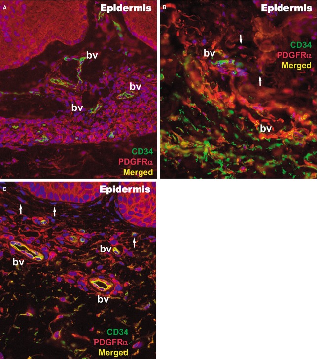
Figure 9.
Immunofluorescence of the papillary dermis for CD34 (green), PDGFRα (red), and CD34/PDGFRα (yellow) in a psoriatic plaque (A), distant uninvolved skin (B), and treated skin (C). Telocytes (white arrows) are double positive for CD34/PDGFRα and have yellow silhouettes at 200× magnification. bv: blood vessels.

