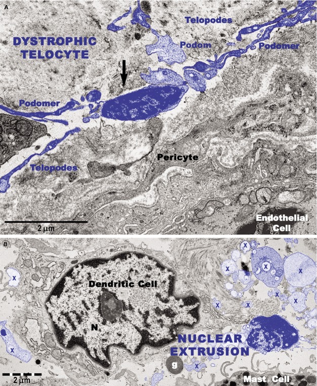
Figure 12.
Transmission electron microscope images show degenerative changes in telocytes (digitally coloured in blue) from a psoriatic plaque. (A) A telocyte with shrivelled nucleus and detached telopodes. The arrow indicates dissolution of the cellular membrane and the cytoplasmic content surrounding the nucleus. (B) An extruded nucleus and cytoplasmic fragments (X) of a telocyte are visible in the vicinity of a dendritic cell. g: granule (of a mast cell).

