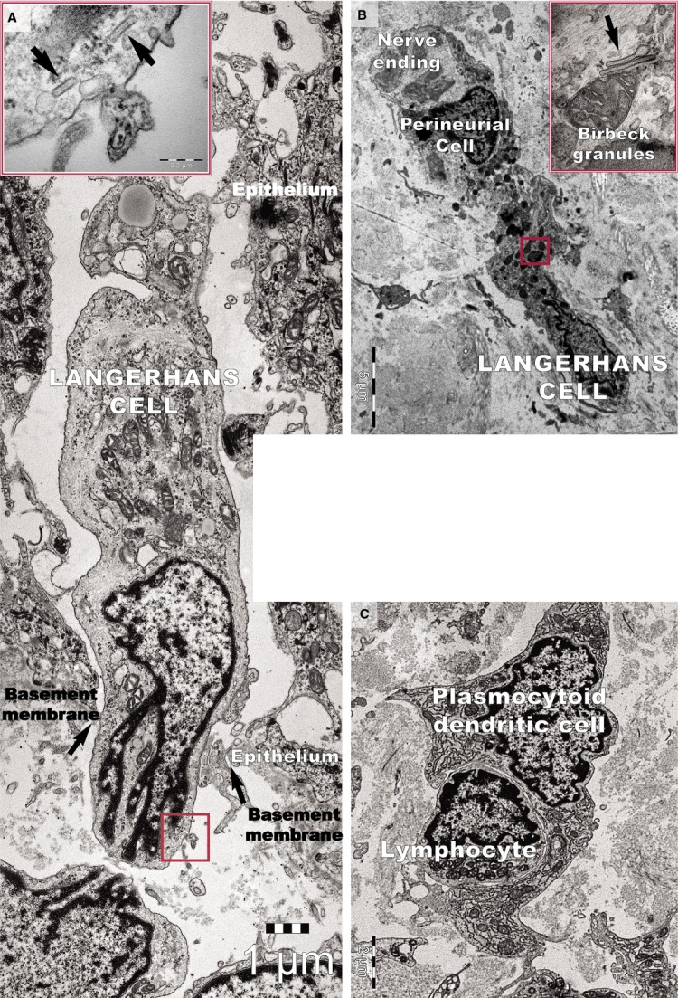
Figure 13.

Transmission electron microscope image of a psoriatic plaque shows (A) a Langerhans cell migrating from the epidermis to the dermis through a gap (arrows) in the basement membrane. The inset shows a higher magnification of the rectangular area, revealing rod-shaped Birbeck granules (arrows). (B) A Langerhans cell from a psoriatic plaque located in the papillary dermis has a cytoplasm filled with lysosomes. The inset shows a higher magnification of the rectangular area, revealing the characteristic Birbeck granules. (C) A plasmacytoid dendritic cell surrounds a lymphocyte in the papillary dermis of a psoriatic plaque. Note the well-developed rough endoplasmic reticulum.
