
Figure 14.
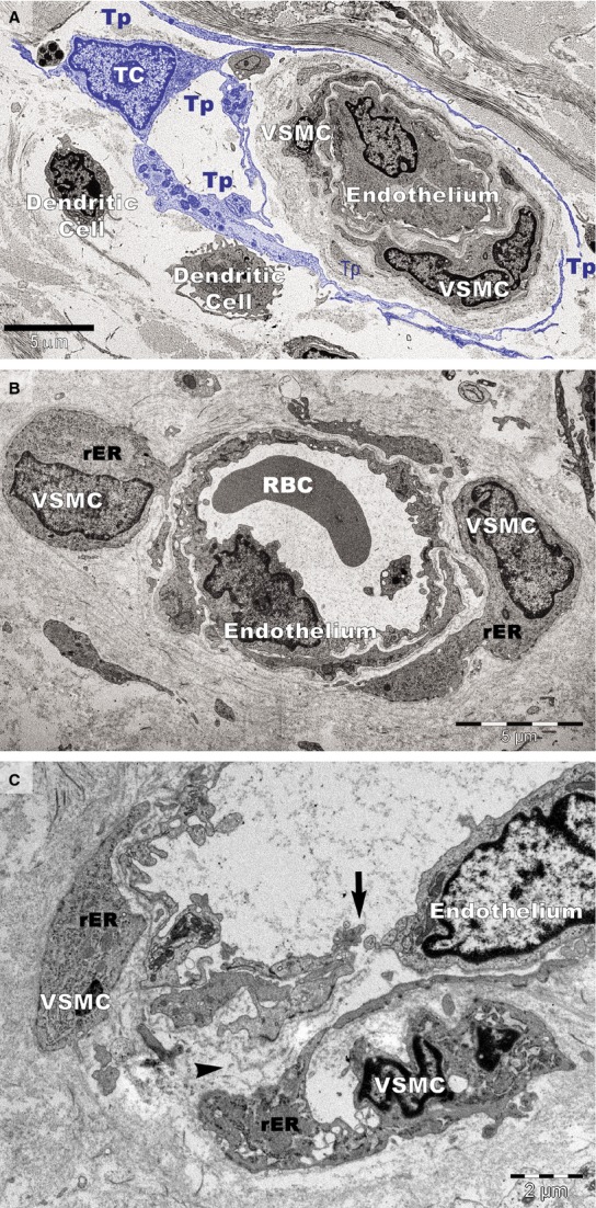
Transmission electron microscopy of small blood vessels in psoriatic plaques. (A) Vascular smooth muscle cell (VSMC) of contractile phenotype is visible in a terminal arteriole surrounded by the telopodes (Tp) of a telocyte (TC; digitally coloured in blue). Dendritic cells have short processes. (B) VSMCs with abundant rough endoplasmic reticulum (rER) in the cytoplasm are bulging into the perivascular space (hypertrophy). RBC: red blood cell. (C) VSMCs of synthetic phenotype in a terminal arteriole are detached from the endothelial layer. A gap (arrow) is visible between endothelial cells that lost their junctions. The basement membrane of the endothelium is also discontinuous (arrowhead).