
Figure 3.
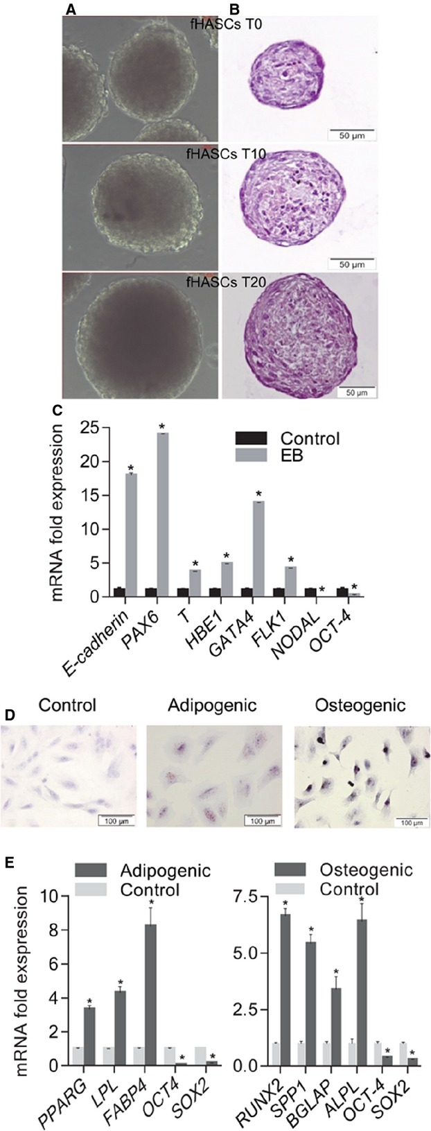
Differentiation potential HASCs. (A) Representative pictures of embryoid bodies (EBs) formed by HASCs in hanging-drop cultures. fHASC-derived EBs that were capable of sustained growth in vitro (T0, 4 days; T10, 10 days; T20, 20 days). (B) EBs stained with haematoxylin/eosin. EB images were obtained by phase-contrast microscopy (×20 magnification). (C) Marker expression before and after EB formation as assessed by RT-PCR (normalized to β-actin). (D) Representative pictures of fHASC differentiation into adipocytes and osteocytes respectively. Differentiation was monitored with Sudan III staining of lipid droplets in terminal adipocyte differentiation and von Kossa staining of calcium deposition in the extracellular matrix in terminal osteoblast differentiation; nuclear staining was with Mayer’s haematoxylin. (E) qRT-PCR analysis of mRNA levels of specific adipogenic (PPARG, LPL, FABP4) and osteogenic (ALPL, OCN, OPN, RUNX2) markers for fHASCs). Analysis was performed at day 21 of differentiation as compared to day 0, which corresponds to fold change = 1. All data shown are representative of three independent experiments conducted on five fHASC lines (*P < 0.05).