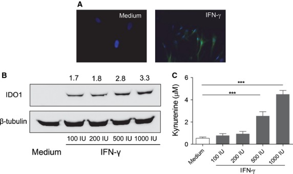
Figure 4.

fHASCs express immunoregulatory IDO1. (A) IDO1 detection by immunofluorescence staining in fHASCs that were treated or not with 1000 U/ml IFN-γ for 24 hrs. Nuclei were counterstained with DAPI (blue) and IDO1 was revealed using a fluorescently labelled secondary antibody. Data are representative of one of three independent experiments using five fHASC lines. (B) Western blot analysis of IDO1 protein expression in fHASCs treated or not with 100, 200, 500 or 1000 U/ml IFN-γ for 24 hrs; β-tubulin was used as a control. (C) IDO1 enzymatic activity was measured by HPLC quantification of tryptophan conversion into kynurenine. Data are representative of three independent experiments involving five fHASC lines ***P<0.05–0.01.
