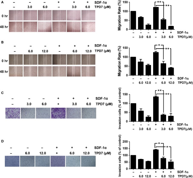
Figure 5.
TPD7 suppressed migration and invasion of breast tumour cells. (A and B) Photographs and quantification of wound of cells treated with TPD7. Wound healing assay was performed for evaluating the inhibitory effect of TPD7 on MDA-MB-435s (A) and MDA-MB-231 (B) cell migration. Confluent monolayers of cells were scarred, and pre-treatment with TPD7 for 48 hrs before being exposed to 100 ng/ml SDF-1α for 24 hrs. The average distance of migrating cells into the wound surface were determined under an inverted microscope. The representative photographs showed the same area at time zero and after 48 hrs of incubation. (C and D) Photographs and quantification of the cell invasion through the Matrigel-coated polycarbonate membrane stained by 0.2% crystal violet. MDA-MB-435s (C) and MDA-MB-231 (D) cells were seeded in the top-chamber of the Matrigel. After pre-treatment with or without TPD7 for 48 hrs, Millicell chambers were then incubated with either the basal medium only or basal medium containing 100 ng/ml SDF-1α for 24 hrs. After incubation, they were assessed for cell invasion as described in Materials and methods. The representative photographs showed the cell migration through the polycarbonate membrane stained by 0.2% crystal violet. The results shown were representative of three independent experiments. *P < 0.05; **P < 0.01 compared with SDF-1α treated control cells.

