Figure 3.

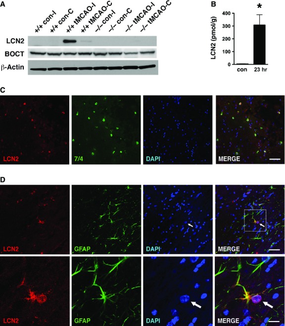
Detection of LCN2 in infiltrating neutrophils and astrocytes in the ipsilateral hemisphere after tMCAO. (A) The ipsilateral (I) and contralateral (C) hemispheres of Lcn2+/+ and Lcn2−/− mice were isolated after 1 hr of tMCAO and 23 hrs of reperfusion. The hemispheres of mice without tMCAO were used as controls (con). The brain homogenates were analysed by western blotting using anti-LCN2 and anti-BOCT antibodies. β-Actin was used as a loading control. (B) The levels of LCN2 in the brain homogenates of ipsilateral hemispheres at 23 hrs after tMCAO were determined by ELISA. The ipsilateral hemispheres of mice without tMCAO were prepared as controls (con). *P < 0.05 compared with control (two-tailed, unpaired t-test). (C) Immunoreactivities of LCN2 (red) and neutrophil-specific marker 7/4 (green) in the ipsilateral hemispheres at 23 hrs after tMCAO were detected using confocal microscopy. (D) Confocal images of LCN2 (red) and GFAP-positive astrocytes (green) in the ipsilateral hemispheres at 23 hrs after tMCAO. Amplified images of LCN2- and GFAP- positive astrocytes are shown in the bottom panels. The arrows indicate the enlarged and eccentric nuclei of GFAP-positive astrocytes. Merged images indicate colocalization of LCN2 with 7/4 or GFAP in yellow. Scale bars, 50 μm (C and D top panel) and 12.5 μm (D bottom panel).
