
Figure 5.
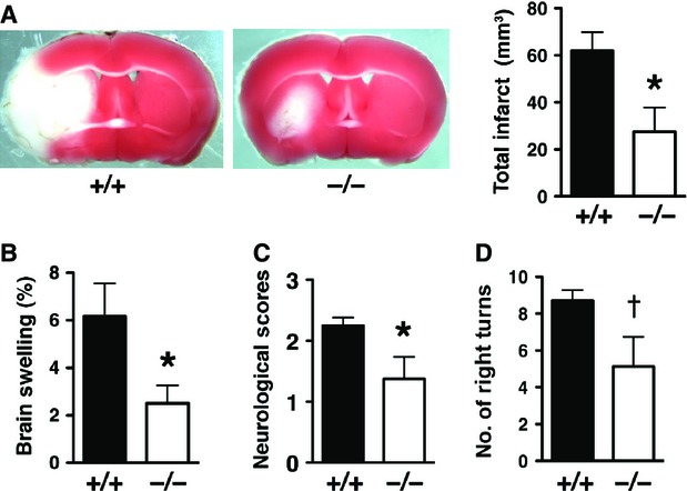
Stroke injury is reduced in LCN2 null mice after tMCAO. (A) Representative images of TTC-stained brain slices from Lcn2+/+ and Lcn2−/− mice after 1 hr of tMCAO and 23 hrs of reperfusion. Viable tissue is stained in red colour, while the infarcted area in white colour. Total Infarct volume (A), brain swelling (B), neurological deficit scores (C) and corner tests (D) were determined after 1 hr of tMCAO and 23 hrs of reperfusion from Lcn2+/+ (n = 8) and Lcn2−/− (n = 8) mice. *P < 0.05 compared with WT mice (two-tailed, unpaired t-test). †P < 0.05 compared with WT mice (one-tailed, unpaired t-test).