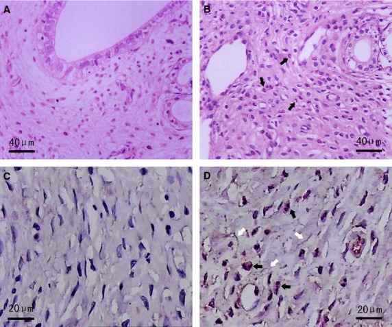
Figure 1.

Haematoxylin and eosin and CD177 IHC staining in AS-affected and -unaffected oviduct tissues. (A) Normal oviduct tissues from the sham group displayed no obvious changes (haematoxylin and eosin staining). (B) Representative microphotographs of acute inflammation in AS-affected oviduct tissues, including SMCs and capillaries swelling, inflammatory congestion and exudation, interstitial oedema and infiltration of neutrophils (black arrows) (haematoxylin and eosin staining). (C) Totally negative CD177 immunostaining indicating no obvious infiltration of inflammatory cells in sham group. (D) Extensive infiltration of neutrophils (black arrows) as indicated by strong positive CD177 immunostaining, together with interstitial fibrosis (white arrows) in AS-affected oviduct tissues.
