
Figure 3.
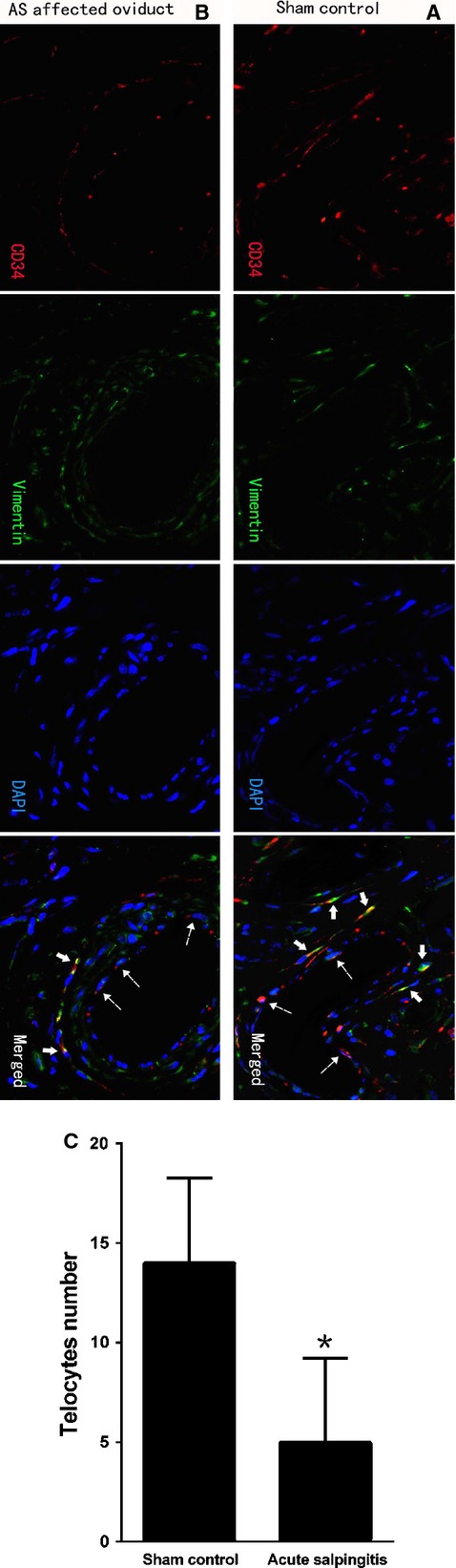
TCs immunodiagnostics by double-labelled immunofluorescence. Dotted arrows indicated CD34-positive vascular endothelial cells. Negative c-kit staining was not shown here; scale bar = 20 μm. (A) CD34 (red) in moniliform cells overlying vimentin (green) cells with DAPI counterstaining (blue) in sham control (solid arrows), indicated the existence of perivascular TCs with special immunophenotype of CD34/vimentin double-positive. (B and C) CD34/vimentin double-positive cells with specific TCs morphology and well-defined nuclei was significantly less densely stained, reduced, sparse or completely absent (solid arrows) in AS-affected oviduct tissues. A statistically significant decrease in the mean number of TCs occurred (P = 0.000). *P < 0.05 versus sham control; error bars = SD.