
Figure 5.
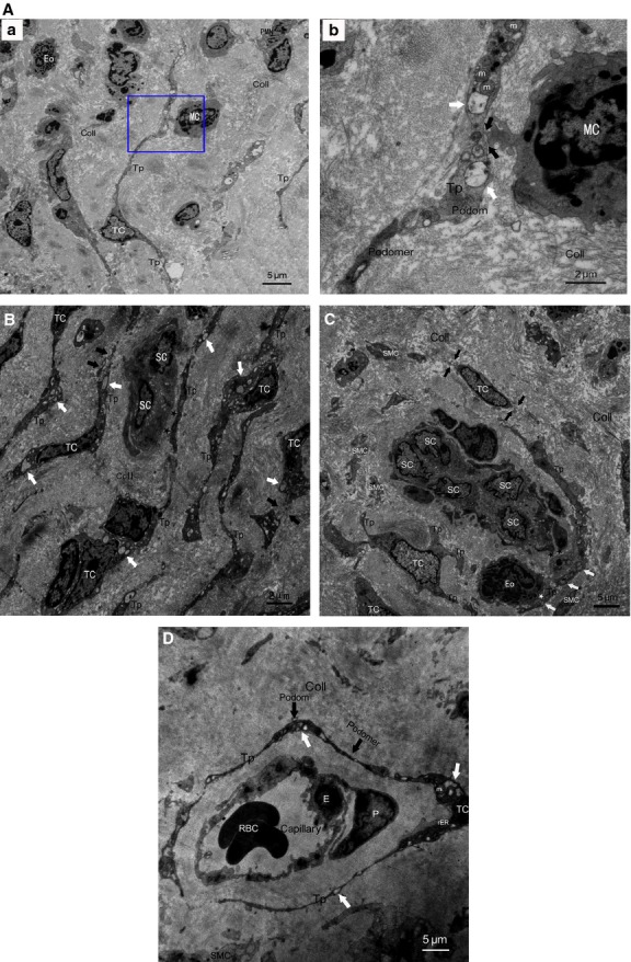
TCs and Tps damage in AS-affected oviduct tissues, accompanied by excessive amount of collagen fibres (Coll) and tissue fibrosis. (A) Intercellular connection between damaged TCs and activated mononuclear cells (MC). (a) Degenerated Tps established closed contact to activated MC which contained dense secretory granules, together with granulocyte infiltration, mainly eosinophils (Eo) and neutrophils (PMN). b higher magnification of the boxed area; (b) synapse (black arrows) between activated MC and degenerated Tps, which contained lots of swollen mitochondria (m) and vacuoles (white arrows), thus indicating degeneration, functional insufficiency of TCs and involvement of TCs in local immunoregulation. (B) Degeneration, discontinue or dissolution of TCs and Tps (black arrows), with cytoplasmic vacuolization (white arrows), accompanied by nearly normal scattered putative stem cells (SCs). Intercellular contacts between TCs and SCs was getting wider or disappeared (black asterisks). (C) Disrupted TC-SC niches which composed of a group of damaged Tps and putative SCs in myosalpinx, with heterocellular contacts getting wider or disappeared between Tps and SCs (black asterisks), Tps and activated Eosinophils (Eo) (white asterisk) with dense secretory granules respectively. Degeneration, discontinue or dissolution of TCs and Tps (black arrows), with swollen mitochondria (m), cytoplasmic vacuolization (white arrows) in Tps, swollen and dissolution of SMCs can be observed. (D) Severely damaged perivascular TCs and Tps, with swollen mitochondria (m), rough endoplasmic reticulum (rER) dilatation and cytoplasmic vacuolization (white arrows), together with damaged endothelial cell (E) and pericytes (P).