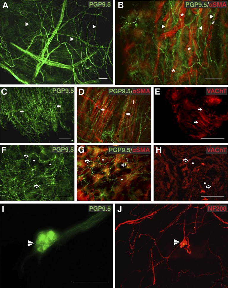
Figure 1.
Autonomic fibers in the human airway mucosa. (A and B) Protein gene product 9.5 (PGP9.5) immunopositive (green, Alexa Fluor 488) subepithelial fibers (arrowheads) were associated with α-smooth muscle actin (SMA)-immunoreactive (red, Alexa Fluor 568) blood vessels (asterisks). (C–E) PGP9.5-immunopositive (green, Alexa Fluor 488) convoluted fibers (arrows) were observed in parallel to bronchial smooth muscle (daggers) labeled with α-SMA (red, Alexa Fluor 568) and a proportion were immunopositive for vesicular acetylcholine transporter (VAChT) (red, Alexa Fluor 568). (F–H) The distinctive circular pattern of PGP9.5 and VAChT immunoreactive fibers (open arrows) around acinar cells of bronchial mucosal glands observed with α-SMA (circles). (I and J) Airway intrinsic autonomic ganglia (split arrowheads) immunoreactive for both PGP9.5 (green, Alexa Fluor 488) and 200-kD neurofilament (NF200; red, Alexa Fluor 568) were observed. Biopsies were from right middle lobe entrance (A, B, E, F, and H), right lower lobe (C, D, and G), right upper lobe entrance (I), and right main bronchus (J). Results are independent experiments of staining in nine biopsies from seven patients. Scale bars = 100 μm. Unprocessed and single-channel images are shown in Figures E1 and E2 with additional images in Figure E3. A video of additional images of separate focal planes from B is shown in Video E1.

