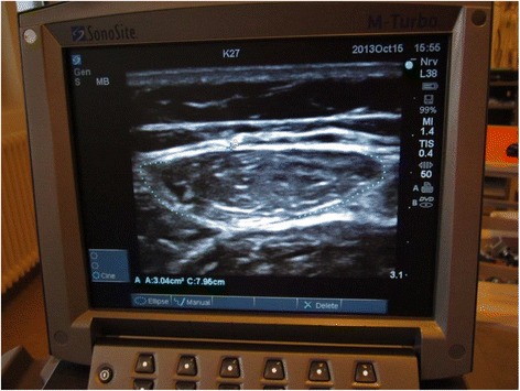
Fig. 2.

Ultrasonographic measurement of the rectus femoris muscle cross-sectional area. The rectus femoris muscle is part of the quadriceps muscle. Patients will be placed in supine position with their back raised to 45 degrees, with their legs in passive extension. The transducer will be placed over the rectus femoris muscle, perpendicular to the long axis of the right thigh, not depressing the dermal surface. Measurements will be made at 2/3 of the distance from the anterior superior iliac spine to the superior patellar border. This distance will be defined when the patient is placed as noted above, not with the patient standing up, since this changes the distance. For the scan, a linear transducer will be used, flat footprint, 5–8 MHz. The muscle is identified visually and an ultrasonographic picture is taken. Using the ultrasonographic software, the outer edge of the muscle is marked, and the cross-sectional area is calculated using planimetry
