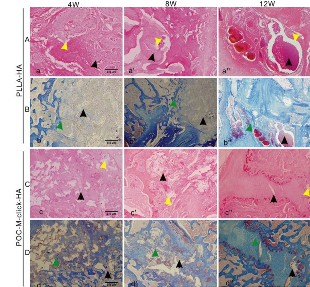
Figure 7.
Photomicrographs of H&E-stained (A and C) and Masson’s trichrome-stained (B and D) tissue sections of the explanted PLLA-HA (A and B) and POC-M-click-HA scaffolds (C and D) 4, 8, and 12 weeks after operation. Yellow arrows indicate vacuoles formed upon material degradation and black arrows indicate the residual unabsorbed materials. Green arrows indicate a large amount of new bone formation surrounding the materials.

