
Fig. 1.
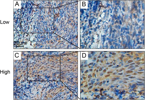
Immunohistochemistry staining of BRCC3 expression in nasopharyn-geal carcinoma tissues. BRCC3 expression was mainly localized within the nuclei of cancer cells. a Tumor with low BRCC3 level (200×); c Tumor with high BRCC3 level (200×); b and d demonstrated the higher magnification (400×) from the area of the box in (a) and (c) respectively. Low: low BRCC3 expression; High: high BRCC3 expression