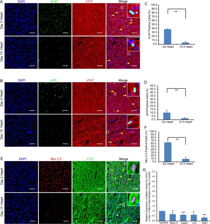
Fig 2. Extent of cardiomyocyte proliferation and differentiation in 2- and 13-day-old mouse hearts.
Immunofluorescent staining showing the presence of proliferating Ki-67+ (A) and mitotic pH3+ (B) cTnT+ cardiomyocytes (yellow arrow heads) at day 2 and day 13. (E) showing the presence of Nkx2.5+ cardiac progenitor cells at day 2 and day 13. Bar charts showing the percentage of cardiomyocytes that are Ki-67+ (C), pH3+ (D) or Nkx2.5+ (F) in 2- and 13-day-old hearts. The data is presented as means ± SD; Scale bar: 50μm, Significantly different: **P<0.01. (G) RT-qPCR assay confirms the immunofluorescent staining that Ki-67+ and pH3 expressions are down-regulated in 13-day-old heart. Relative expression values are presented as means ± SD. Significantly different: *P<0.05, **P<0.01.

