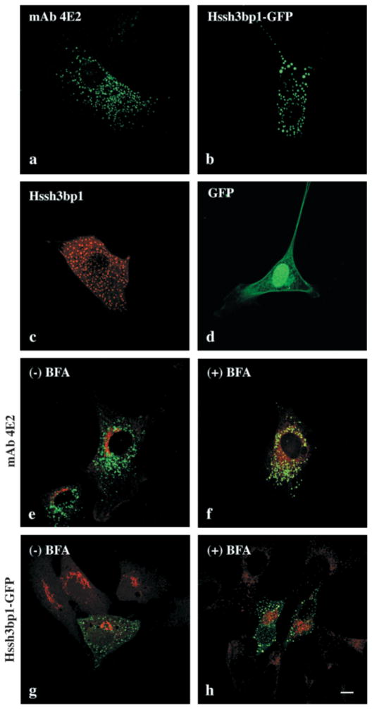
Fig. 1.
Association of Hssh3bp1 and Hssh3bp1-GFP with vesicles resistant to BFA treatment in NIH3T3 cells. (a) Nontransfected NIH 3T3 cell stained with mAb 4E2. (b) Hssh3bp1-GFP-transfected cell. (c) Hssh3bp1-transfected cell stained with mAb 4E2. (d) a cell transfected with GFP alone. (e and f) Cells costained with mAb 4E2 (green) and antibody to mannosidase II (red); (g and h) cells transfected with Hssh3bp1-GFP (green) and costained with antibody to mannosidase II (red). (e and g, f and h) Cells before and after BFA treatment, respectively. Bar, 10 μm.

