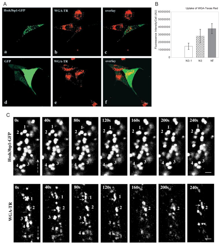
Fig. 6.
Effect of Hssh3bp1-GFP overexpression on endocytosis of WGA-TR in NIH 3T3 fibroblasts. (A) Confocal images were obtained from cells expressing Hssh3bp1-GFP (a and b), or GFP only (d and e). (a and d) Green fluorescence of GFP proteins; (b and e) red fluorescence of WGA-TR. (a and b, d and e) Represent confocal images from the same cells, respectively; (c) an overlay of a and b images; (f) an overlay of d and e images. Note that the cell expressing Hssh3bp1-GFP (the cell in the center of b), endocytosed less fluorescence than nonntransfected cells (cells at the top of b and in the left of b). Bar, 10 μm. (B) Measurements of endocytosed fluorescence in cells transfected with Hssh3bp1-GFP (N3-1), with GFP only (N3), and nonntransfected (NT). In each group not less than 20 individual cells were analyzed (bar represents mean ± s.d.). Similar results were obtained in three independent experiments. (C) Sequential confocal images from a live cell transfected with Hssh3bp1-GFP (upper plates) or from a nontransfected cell (WGA-TR) (lower plates) following uptake of WGA-TR. Images were captured every 40 seconds for 240 seconds. Hssh3bp1-GFP plates: Vesicles 1 and 2, and vesicles 3 and 4, move towards each other, their images overlap, and then they separate. WGA-TR plates: note formation of short tubules from irregularly shaped structures 1 and 3 (compare 1 at 0 seconds and 40 seconds, and 3 at 40 seconds and 80 seconds). Some possible budding or fusion events are indicated by short arrows. Emergence of peripheral structures which then move towards structure(s) located closer to perinuclear area is common (see structures located above vesicles 1, frames 40 seconds through 120 seconds, and vesicle 2, frames 160 seconds through 240 seconds). Vesicle 4 moves downwards in first four frames. Vertical arrows indicate direction towards perinuclear area of cells. Note substantial morphological changes of WGA-TR vesicles in subsequent frames in comparison to little changes in Hssh3bp1-GFP vesicles. Bar, 2 μm.

