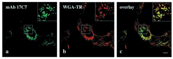
Fig. 8.

mAb 17C7 to the erythroid α-spectrin SH3 domain stains macropinocytic vesicles in NIH 3T3 cells. (a–c) Cells were allowed to endocytose WGA-TR (red) for 5 minutes and stained with mAb 17C7, (green); (c) an overlay of a and b images. Boxed areas in upper right (a–c) of each panel show enlargements of indicated areas. Bar, 10 μm.
