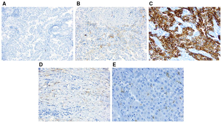
FIG. 1.
CD10 immunohistochemical analysis using a tissue micro-array (original magnification, ×200). a Tumor cells are negative for CD10; b tumor cytoplasm is weakly positive for CD10; c tumor cytoplasm is strongly positive for CD10; d tumor-related stroma is positive for CD10; e granulocytes infiltrating in tumor cells are positive for CD10

