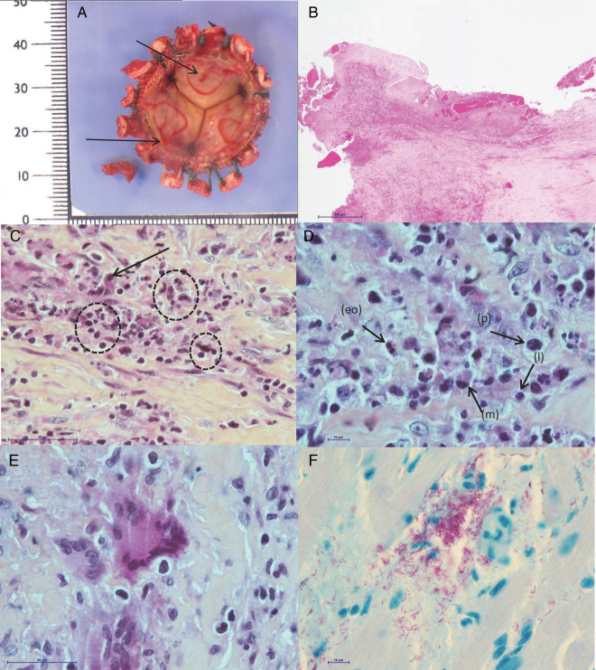
Figure 2.
Histological examinations and Ziehl-Neelsen staining of explanted bioprostheses. A, Macroscopic aspect. Endothelial ulcerations (black arrows) with fibrin replacement. B, Vegetation with leukocytes and fibrin matrix at the surface of the prosthesis (hematoxylin-eosin staining) (scale bar = 500 μm) (low magnification). C, Enlargement of (B): focus on the inflammatory infiltrate with macrophages (black arrow) and foci of necrotic neutrophils (dotted circles) (scale bar = 50 μm). D, Enlargement of (C): focus on plasma cells (p), lymphocytes (l), macrophages (m), and rare eosinophils (eo) (scale bar = 10 μm). E, Enlargement of (C): focus on few scattered multinucleated giant cells (scale bar = 50 μm). F, Ziehl-Neelsen staining revealing numerous acid-fast bacilli (in pink) (scale bar = 10 μm).

