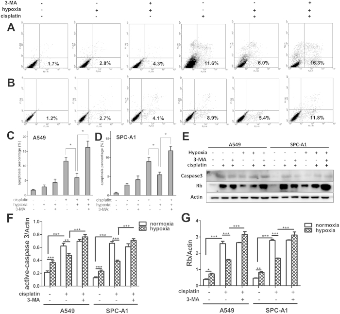
Figure 4. 3-MA completely abolished the effect of hypoxia on cisplatin-induced apoptosis.
A549 (A and C) and SPC-A1 (B and D) cells were treated with or without cisplatin for 12 h under normoxia or hypoxia, followed by Annexin-V and PI staining and FACS analysis. Cells represented early apoptosis (Annexin-V+/PI−) were calculated (C and D). (E) The expressions of activated-caspase-3 and Rb were detected by western blot. (F) The relative expression of activated-caspase-3. (G) The relative expression of Rb. Each band was quantified using densitometry. Data were shown as the means ± SD. *p < 0.05, **p < 0.01, ***p < 0.001. All studies were representative of at least three independent experiments. Blot images were cropped for comparison.

