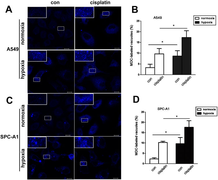
Figure 5. Hypoxia up-regulated the number of autophagolysosome induced by cisplatin.
A549 (A, upper) and SPC-A1 (C, lower) cells were treated with cisplatin for 12 h under normoxia or hypoxia respectively. MDC was loaded into cells and incubated at 37 °C for 15 min, then visualized using confocal laser scanning (400×). The ratio of A549 or SPC-A1 cells containing MDC-labeled vacuoles was calculated and showed in (B and D). The magnified representative vacuoles were showed upper left. Data were shown as the means ± SD. *p < 0.05.

