
Figure 2.
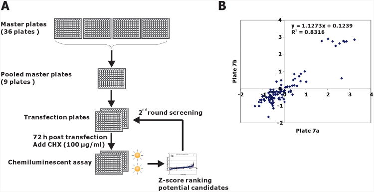
Chemiluminescence assay of PL-NKX3.1 degradation in 96-well plates. A. Schematic diagram of the siRNA strategy for kinome siRNA screening. From master plates, four target-specific siRNAs were pooled into working transfection plates, siRNA pools from each transfection plate were reverse transfected into LNCaP-PLNKX7 cells. After 72 h of transfection, the plates were treated with cycloheximide for 1 hr. Chemiluminescent assay was performed with Prolabel Detection Kit II. B. Plate-to-plate reproducibility is shown in a pairwise plot of Z-score values from replicate plates 7a and 7b that were the master plates used for first round screening. Cells were plated in a replicate manner, but the transfections were performed separately at different times. Linear regression indices of the pairwise comparison are shown.