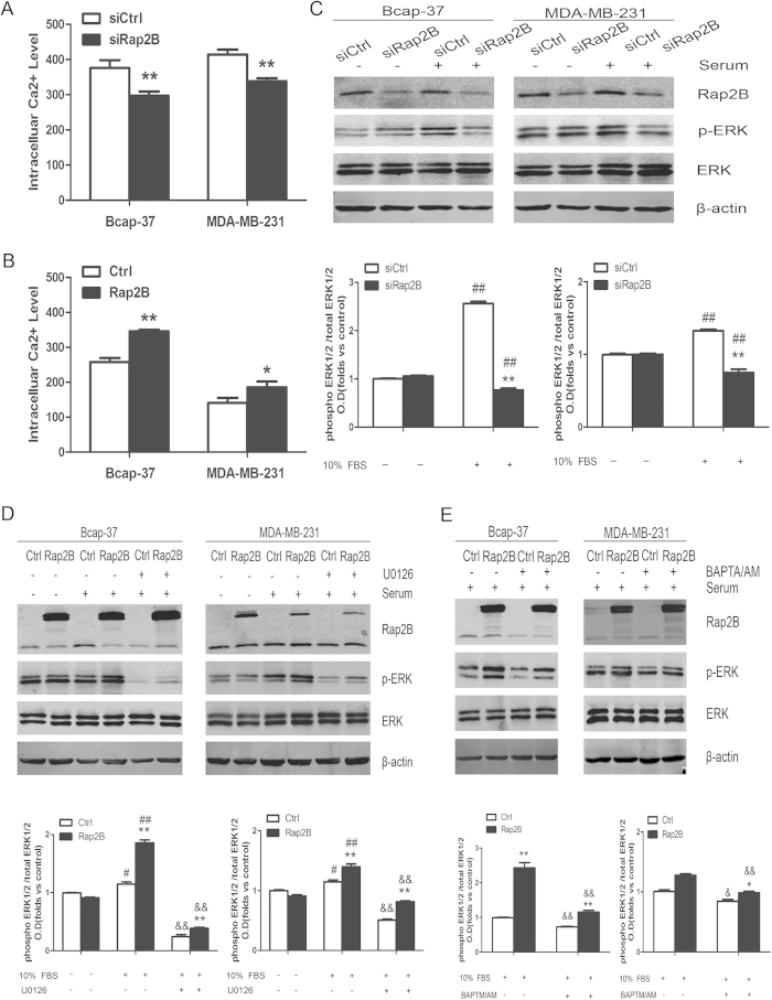
Figure 3. Rap2B can increase the intracelluar calcium level and induce ERK1/2 phosphorylation.
(A) Rap2B siRNA could decrease intracelluar calcium level in Bcap-37 and MDA-MB-231 cell lines, as measured by a flow cytometer. (B) Rap2B overexpression increased intracelluar calcium level in breast cancer cells. (C) After breast cancer cells were transfected and starved, some cells were treated with 10% FBS and some were not. Western blot analysis of the protein levels of Rap2B, p-ERK, and ERK in Rap2B knockdown and control siRNA group for both cell lines. (D) After transfection and starvation, cells were incubated in the presence or absence of U0126 for 30 min. Then, some cells were treated with 10% FBS and some were not. Western blot analysis of the protein levels of Rap2B, p-ERK, and ERK in Rap2B overexpression and control group for both cell lines. (E) After transfection and starvation, cells were incubated in the presence or absence of BAPTA/AM for 30 min. Then, some cells were treated with 10% FBS. Western blot analysis of the protein levels of Rap2B, p-ERK, and ERK in Rap2B over-expression and control group for both cell lines. All experiments were carried out in triplicate. Data are presented as mean ± SD (n = 3). *P < 0.05, **P < 0.01 in comparison to respective group, #P < 0.05, ##P < 0.01 in comparison with respective 10% FBS-untreated group, and &P < 0.05, &&P < 0.01 in comparison with respective inhibitor-untreated group.

