Fig. 1.

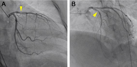
Coronary angiography. a. Right anterior oblique and caudal view. b. Left anterior oblique and cranial view. A stent thrombosis is visible (arrowheads) in the in-stent segment of the proximal left anterior descending artery

Coronary angiography. a. Right anterior oblique and caudal view. b. Left anterior oblique and cranial view. A stent thrombosis is visible (arrowheads) in the in-stent segment of the proximal left anterior descending artery