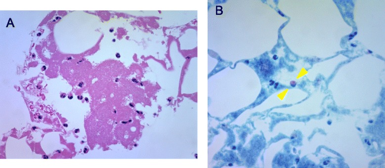
Fig. 4.

Histological examination of aspirated thrombi. a. High-power (original magnification 400×) microphotograph showing the infiltration of inflammatory cells in aspirated fibrin-rich thrombus as observed after haematoxylin and eosin staining. b. High-power (original magnification 400×) microphotograph showing the eosinophilic infiltration as observed after Giemsa staining (between arrowheads)
