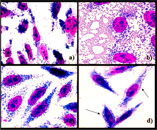
Figure 1.
Micrographs of adherence patterns on HEp2 cells of Escherichia coli obtained from faeces of children with diarrhoea and controls from Porto Velho-RO, Brazil: a) LA pattern after 3 h of incubation with typical EPEC; b) AA pattern after 3 h of incubation with EAEC; c) DA pattern after 3 h of incubation with DAEC; d) LAL pattern after 6 h of incubation with atypical EPEC (arrows).

