. 2015 Mar 31;46(1):207–217. doi: 10.1590/S1517-838246120130859

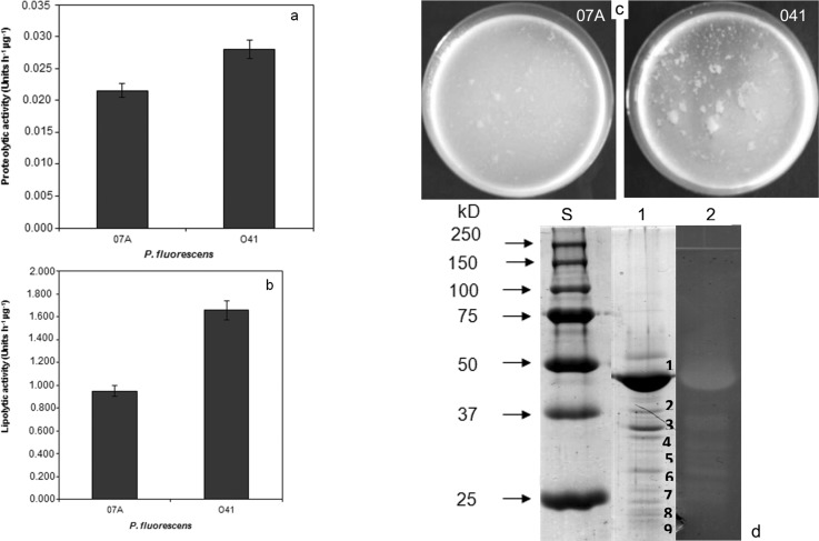
Figure 1. Production of extracellular hydrolytic enzymes by P. fluorescens . A: Proteolytic activity in the supernatant of TYEP medium; B: Lipolytic activity in the supernatant of TYEP medium; C: Samples of reconstituted skin milk powder (12%) inoculated with P. fluorescens 07A and 041 after 18 h of incubation at 25 °C. Data represent the average of duplicate experiments; D: Coomassie-stained SDS-PAGE and azocasein zymogram on 12% PAA-gels visualizing protease production by P. fluorescens grown in TYEP medium supplemented with 0.25% CaCl 2 . Lanes S: molar mass standard (Biorad); lane 1: SDS-PAGE of ammonium sulfate precipitated proteins of P. fluorescens 041 supernatant; lane 2: azocasein zymogram of ammonium sulfate precipitated proteins of P. fluorescens 041 after proteins precipitation with ammonium sulfate.

Figure 1