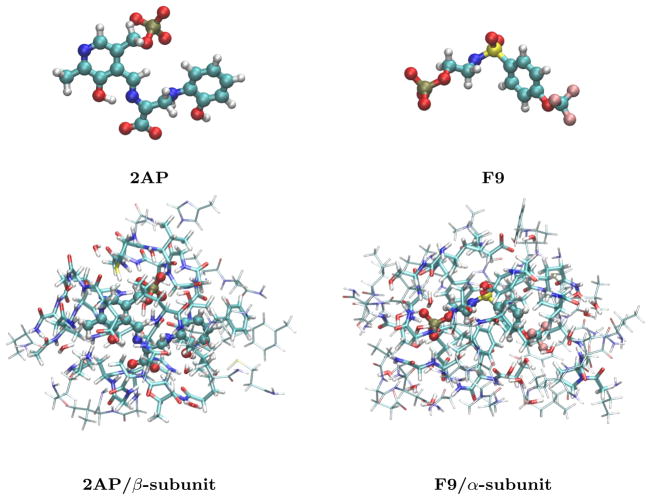
Fig. 1.
The bare 2AP quinonoid substrate-coenzyme complex and F9 ligand (top) and their structures bound in the β and α subunits of tryptophan synthase, respectively. The bottom structures indicate the 5 Å clusters (cylinders) and the larger 7–8 Å clusters (wireframe) extracted from tryptophan synthase.

