Figure 3.

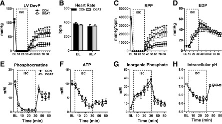
Improved recovery after low-flow ischemia in MHC-DGAT1 hearts. A–D: LVDevP, heart rate, RPP, and LV end diastolic pressure (EDP) in perfused hearts during 30 min of low-flow ischemia (ISC) (1% of baseline [BL]) followed by 55 min of reperfusion (REP). Hearts were perfused with a mixed substrate buffer consisting of 5.5 mmol/L glucose, 0.4 mmol/L LCFAs, 1.2 mmol/L lactate, and 50 μU/mL insulin (n = 7–8 at each time point). E–G: PCr, ATP, and Pi content measured with 31P NMR spectroscopy in isolated perfused hearts (n = 5 for each group at each time point). H: Intracellular pH assessed by 31P NMR spectroscopy in isolated perfused hearts, calculated by the chemical shift between Pi and PCr (n = 5 each group). *P < 0.05 vs. CON.
