Figure 7.

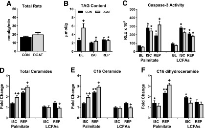
TAG incorporation, lipolysis, and ceramide accumulation in MHC-DGAT1 hearts perfused with palmitate. A: Incorporation rate of palmitate into the TAG pool during the reperfusion period in isolated perfused hearts (n = 6–7). B: TAG content measured in lipid extracts from perfused hearts freeze-clamped at the end of baseline (BL), ischemia (ISC), or reperfusion (REP) (n = 5–8 per group at each time point). C: Caspase-3 activity measured in tissue lysates from isolated hearts perfused with palmitate or LCFAs at the end of baseline, ischemia, and reperfusion. Values shown are relative light units (RLU × 103; n = 3–6 at each time point). D–F: Total abundance of ceramides, palmitoyl-ceramide (C16 ceramide), and palmitoyl-dihydroceramide (C16 dihydroceramide) in isolated hearts perfused with palmitate or LCFAs at the end of ISC and REP. Values are expressed as fold change relative to CON hearts at baseline (n = 4–5 at each time point). *P < 0.05 vs. baseline; **P < 0.05 vs. ischemia; +P < 0.05 vs. CON at same time point.
