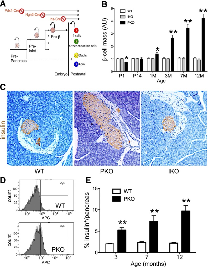
Figure 1.
Increased β-cell mass following pan-pancreatic Foxo1 ablation. A: Strategy to delete Foxo1 in all pancreatic cell types, endocrine progenitors, and differentiated β-cells. B: Analysis of β-cell mass by immunohistochemistry (n = 6 each genotype and each age). At each time point, β-cell mass in WT littermates was normalized to 1 for clarity. C: Representative images of insulin immunohistochemistry (brown) of pancreatic sections from 3-month-old mice used for the analysis in panel B. Original magnification ×100. Analysis of β-cell number by flow cytometry, showing a representative plot (D) and quantification of results (E). We digested pancreata into single cells and fixed and stained the cells with anti-insulin antibodies. Insulin+ cells were normalized by DNA concentration (n = 6 each genotype). *P < 0.05; **P < 0.01. AU, arbitrary units; M, month; P, postnatal day.

