
Fig. 6.
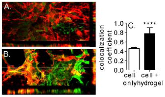
3-dimensional reconstruction of (A) cell only and (B) cell + hydrogel sections stained for GFP-labeled transplanted cells and DOUBLECORTIN. (C) Colocalization analysis shows that the majority of DCX positive signal seen in cell + hydrogel condition is from transplanted cell differentiation.