Fig. 5.

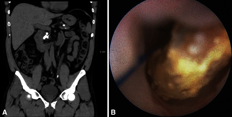
Patient with left ureteral stone and PSS. a Appearance of the stone along the suture in the dilated ureter on CT scan. b No ureteral inflammation is visible in contact with the stone (endoscopic appearance)

Patient with left ureteral stone and PSS. a Appearance of the stone along the suture in the dilated ureter on CT scan. b No ureteral inflammation is visible in contact with the stone (endoscopic appearance)