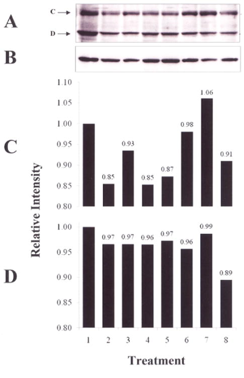
Figure 2.

Western blot of U-STAT1. T/C28a2 chondrocytes were incubated for 0 or 30 min with 1) no additions; 2) DMSO; 3) 4) rhIL-6 (50 ng/ml); 5) rhIL-6+Janex-1 (100 μM) or rhIL-6 plus sIL-6R (30 ng/ml). The western blot shows A) U-STAT1A (band C); U-STAT1B (band D); B) β-actin; D) Quantified U-STAT1A and U-STAT1B. Lane 1, No additions, 0 min; Lane 2, DMSO, 0 min; Lane 3, rhIL-6+Janex-1, 0 min; Lane 4, No additions, 30 min; Lane 5, DMSO, 30 min; Lane 6, rhIL-6+Janex-1, 30 min; Lane 7, rhIL-6, 30 min; Lane 8, rhIL-6+sIL-6R, 30 min.
