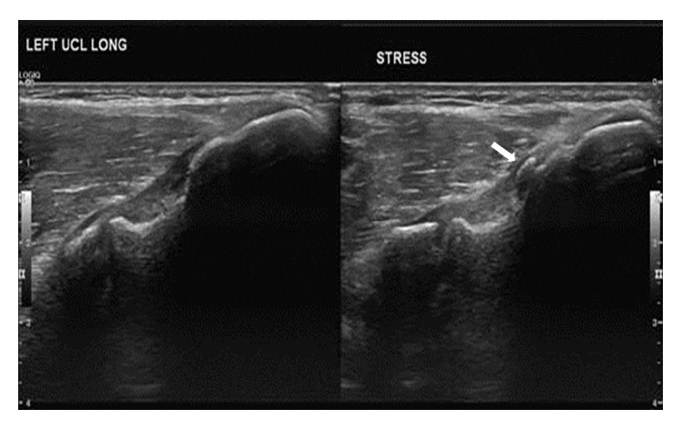
Figure 1.

Preoperative ultrasound. Images demonstrate mild thickening of the proximal fibers of the proximal ulnar collateral ligament (medial epicondyle is to the right of image, and ulna is to the left of the image), likely sequela of prior or chronic repetitive low grade injury. Small area of heterotopic ossification embedded within the proximal fibers of the ulnar collateral ligament (arrow). Dynamic stress images demonstrate mild “gapping” of the medial compartment (2 mm) suggestive of laxity.
