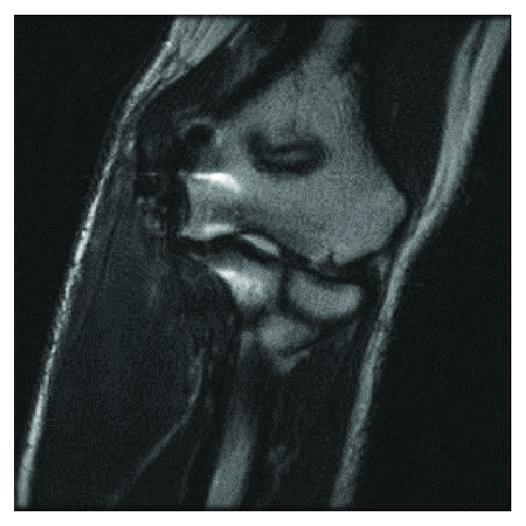
Figure 8.

Three-month postoperative radiograph. Coronal T2 weighted images demonstrate low signal intensity fibers extending from the medial epicondyle of the distal humerus to the medial margin/sublime tubercle of the proximal ulnar. No tear, deformity, or retraction of the ulnar collateral ligament reconstruction site.
