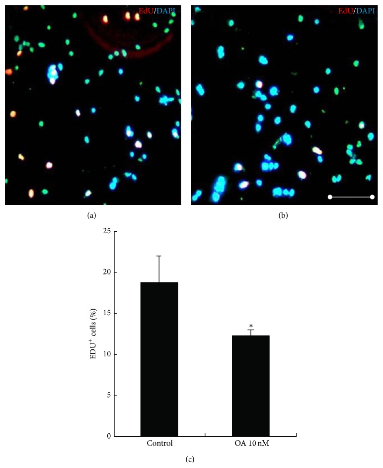
Figure 2.
Effects of OA on the proliferation of NSCs. Single NSCs were plated at a density of 5000 cells per well in PDL-coated 96-well plate for 12 h. Then, cells were subjected to 10 nM EdU for 2 h, followed by addition of 10 nM OA (b) or without added OA (a). Then EdU immunofluorescence analysis was performed. The cell nuclei were counterstained with DAPI. The percentage of EdU-positive cells in a total of 1000 cells was calculated. OA significantly inhibited DNA incorporation (c). Scale bars: 100 μm. Results were expressed as mean ± SD from four independent experiments. * P < 0.05 versus control.

